

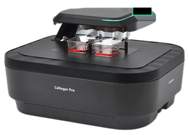
Purchasing imaging equipment for a laboratory or research facility is rarely a straightforward decision. The stakes extend beyond the initial cost. Workflow disruptions, sample integrity, operator dependencies, and long-term data consistency all hinge on whether the equipment selected can actually perform under the demands of real laboratory conditions. When it comes to cell imaging, the gap between a system that meets its specification sheet and one that holds up across daily operations is often significant.
Most procurement decisions for imaging systems happen under pressure — a lab scaling its throughput, a research program shifting toward higher-volume screening, or a facility trying to reduce the manual overhead that comes with traditional microscopy. What gets overlooked in that pressure is the evaluation of features that affect long-term reliability, not just immediate performance. This guide is intended to help scientists, lab directors, and procurement leads think through the right questions before committing to a platform.
Understanding What an Automated Cell Imaging System Actually Does
An automated cell imaging system integrates image acquisition, sample handling, and data processing into a coordinated workflow that minimizes the need for continuous operator input. Unlike conventional microscopy, where technicians manually focus, capture, and annotate images, automated systems run protocols across plates or slides with consistent parameters and no operator variability between samples. The value here is not just speed — it is reproducibility. When you browse options across a range of platforms, such as those listed in this automated cell imaging system catalog, the differences between instruments become clearer in context of application requirements rather than raw specifications.
Understanding what the system is doing at a mechanical and computational level matters because it directly affects what happens when something goes wrong, how the system scales with your work, and how much technical dependency you inherit by choosing a particular platform.
Image Quality and Optical Consistency
The optical performance of an imaging system determines the foundational quality of every downstream analysis. Poor optics produce inconsistent focal depth, uneven illumination, and chromatic aberration — all of which introduce noise into data before any analysis software has touched the image. This is not a problem that software can fix after the fact.
Illumination Uniformity Across the Field
In high-throughput workflows, illumination must remain consistent not just within a single image but across an entire plate run. Uneven field illumination causes intensity gradients that skew quantitative measurements, particularly in fluorescence-based assays where signal intensity is tied directly to biological interpretation. A system that performs well in a single-well demonstration may behave differently when running a full 384-well plate, especially if illumination hardware degrades under thermal load.
Autofocus Reliability
Autofocus is one of the most operationally critical components in any automated imaging platform. Systems that rely on a single focus strategy often struggle with samples that have low contrast, sparse cell density, or variable plate geometry. Hardware-based autofocus, which uses an infrared beam reflected from the coverslip or well bottom, tends to be more stable than software-only contrast detection. The practical implication is that unreliable autofocus causes failed acquisitions — and in time-sensitive assays, a failed run cannot simply be repeated.
Throughput Capacity and Plate Format Flexibility
Throughput is not purely a speed metric. It describes how many samples a system can process without requiring manual intervention, reconfiguration, or operator monitoring. A system with high theoretical throughput but poor plate-handling mechanics will routinely require restarts, adjustments, or manual error recovery — which reduces actual output significantly.
Compatibility with Standard and Non-Standard Formats
Laboratories rarely standardize completely on a single plate format. Workflows evolve, collaborators use different formats, and assay protocols change. A system that handles only common plate formats limits the flexibility of the lab over time. Before purchasing, it is worth confirming how the system behaves with specialty plates, chambered slides, or dishes — and whether those formats require separate calibration files or hardware attachments that add friction to daily operations.
Software Architecture and Data Management
The software layer of an imaging system is where many procurement decisions go wrong. Hardware can be evaluated during a demonstration. Software limitations often only emerge weeks into deployment, once users are working with real datasets under real conditions.
On-Instrument vs. Offline Analysis
Some systems require data to be exported before meaningful analysis can occur. Others support real-time segmentation and quantification during acquisition. The distinction matters operationally because real-time feedback allows runs to be flagged or adjusted before completing a full plate, saving time and sample material. Offline-only systems are not inherently inferior, but they create a processing bottleneck that scales poorly as throughput increases.
Data Export and Integration with External Tools
Proprietary data formats create long-term dependencies. If image data cannot be exported in open formats compatible with third-party analysis platforms, the lab becomes locked into a single vendor’s software roadmap. As described in standards documentation from the U.S. Food and Drug Administration, data interoperability and traceability are increasingly central to research and regulatory workflows. Systems that produce data in formats aligned with these expectations reduce compliance overhead over time.
Environmental and Incubation Controls
Cell biology depends on maintaining stable environmental conditions. Live-cell imaging, in particular, requires precise control of temperature, humidity, and carbon dioxide levels during acquisition. A system without integrated environmental controls forces researchers to pre-equilibrate samples under conditions that may change during a long imaging run — introducing variability that is difficult to detect and impossible to correct retrospectively.
Stability Over Extended Runs
For kinetic studies or long-duration live-cell protocols, the system must maintain environmental stability not just at the start but across hours or days. Thermal drift affects both the biological sample and the optical components. A system that stabilizes quickly but drifts over time will produce data that appears valid at first review but reveals inconsistencies during statistical analysis. This is a particularly costly failure mode in studies where the imaging run itself cannot be repeated due to sample limitations.
Ease of Protocol Development and Validation
A system is only as useful as the speed at which trained users can develop and validate reliable protocols. Platforms with deeply nested software interfaces or limited accessibility for assay customization slow down adoption and increase the risk of protocol errors. The best systems allow users to build, test, and modify acquisition workflows with a reasonable learning curve — without requiring vendor support for routine adjustments.
Maintenance Requirements and Service Support
Instrument downtime in a research or clinical setting has real costs — reagents are wasted, timelines slip, and in some cases, irreplaceable biological samples are lost. Before purchasing, it is worth understanding how the system’s components are rated for maintenance frequency, how quickly replacement parts can be sourced, and what the vendor’s service response model looks like in practice.
Preventive Maintenance Documentation
Systems that come with clear, accessible preventive maintenance schedules allow lab staff to perform routine tasks without scheduling a service visit. This reduces both cost and unplanned downtime. Platforms with opaque maintenance requirements, or those that require specialized technicians for basic servicing, tend to accumulate deferred maintenance — which eventually leads to system failures at the worst possible times.
User Access Controls and Audit Trails
In regulated environments, including pharmaceutical research, diagnostics development, and clinical application, data integrity controls are not optional. The system must support role-based user access, logged acquisition events, and traceable modification history. These are not advanced features — they are baseline requirements for any environment subject to quality standards or audit expectations.
Scalability and Upgrade Path
A system purchased for a single assay type will often be expected to support additional applications over its operational lifetime. The question is whether the platform’s hardware and software architecture can accommodate that expansion without requiring replacement. Modular systems that support additional detection channels, different objective sets, or expanded environmental controls through firmware and hardware upgrades offer substantially more long-term value than closed platforms optimized for a narrow use case.
Vendor Stability and Application Support
The commercial stability of the vendor behind an imaging platform has direct implications for long-term support, software updates, and parts availability. A system from a vendor with limited market presence may perform well initially but become unsupported within a few years — leaving the lab with hardware that cannot be serviced or updated. Application support — not just technical support — is also worth evaluating. Vendors who assign dedicated application scientists to help users develop and troubleshoot assays provide a different level of value than those who offer only reactive helpdesk support.
Concluding Considerations
Selecting an automated cell imaging system is a decision that affects laboratory operations for years. The features described above are not a checklist to complete quickly — they are dimensions of evaluation that require honest assessment against your specific workflow, team capability, data requirements, and long-term research direction.
The systems that perform best over time are rarely those with the most impressive demonstration runs. They are the ones whose optics hold up under sustained use, whose software integrates cleanly with existing data infrastructure, whose maintenance demands fit within the team’s capacity, and whose vendors remain engaged after the purchase is complete. Approaching this evaluation with that long-term perspective — rather than optimizing for initial performance metrics alone — is the most reliable way to make a decision the lab will not regret.
